Pulmonary Atresia
Symptoms will be evident soon after birth - Cristiano Antonino
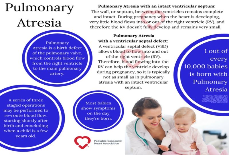
image by: Conquering CHD
HWN Recommends
Prenatal Pathologies, Congenital Heart Defects: Pulmonary Atresia
In intrauterine life, according to the data available to us, this anomaly generally does not impair the child’s development because it is the placenta that supplies the body with the necessary oxygen instead of the lungs. The blood entering the right side of the heart through a hole (foramen ovale) passes from the right atrium to the left atrium, then to the left ventricle and from there it can be pumped to the rest of the body through the aorta.
However, as it cannot reach the lungs via the normal route (right ventricle, then valve and pulmonary artery, then lungs) the blood reaches them via the ductus arteriosus, a vessel that connects the aorta to the pulmonary artery. This structure…
Resources
An Adult with Pulmonary Atresia: Kristen’s Story
There is no cure for congenital heart disease or Pulmonary Atresia, yet. Kristen has undergone four temporary fixes and her scar is a reminder of that. “I embrace my scar because it is a part of who I am. It is a reminder of everything I have been through.
Brooke’s Story
Fast forward a few months to when our cheeky little monkey was born, and we learned that she had a pulmonary atresia with intact ventricular septum (the valve that controls blood flow from the heart to the lungs doesn’t form at all, making it difficult for the blood to get to the lungs to be oxygenated), and narrow, practically non-existent coronary arteries.
Pulmonary Atresia And Pulmonary Stenosis
Pulmonary atresia with a ventricular septal defect is synonymous with an extreme form of Tetralogy of Fallot and Type IV truncus arteriosus or psueudotruncus.
Pulmonary Atresia With Intact Ventricular Septum
The spectrum of this disease range from simple membranous pulmonary atresia (PA) with normal-appearing right ventricle (RV) to hypoplastic RV with abnormal connections between the right ventricle and coronary arteries.
Pulmonary Atresia With Ventricular Septal Defect
Pulmonary atresia with ventricular septal defect (PAVSD) is one of the cyanotic congenital heart diseases. It occurs due to under or maldevelopment of the right ventricular outflow tract along with atresia of the pulmonary valve and trunk. It also involves overriding of the aorta and a large ventricular septal defect. Previously it was termed as truncus arteriosus type IV. Whether it is a different entity or a severe form of Tetralogy of Fallot (TOF) is still controversial, but there is no denying the occurrence of PAVSD.
Smart Cardiology Opinions
Pulmonary Atresia is not a common disorder. Most of the time it is likely to appear with other heart disorders. This problem is said to occur during the early weeks of pregnancy that is, during fetal development.
The Modern Surgical Approach to Pulmonary Atresia with Ventricular Septal Defect and Major Aortopulmonary Collateral Arteries
Pulmonary atresia with ventricular septal defect and major aortopulmonary collateral arteries (PA/VSD/MAPCAs) is a complex congenital heart defect that includes a heterogeneous subgroup of patients characterized by different pulmonary perfusion patterns, of which MAPCAs are an important, although not the only source.
Why patent ductus arteriosus is absent in pulmonary atresia with VSD?
Before answering the above question , there need to be a correction to the question itself . PDA is persistence of ductus arteriosus . In pulmonary atresia ductus itself is not formed .So the question should ideally be Why ductus is absent in pulmonary atresia with VSD. Ductus is formed from the dorsal portion of left 6th arch .The sixth arch also gives raise to right and left pulmonary artery.This can happen only if everything from aortic arch and pulmonary artery development is normal
 Prenatal Pathologies, Congenital Heart Defects: Pulmonary Atresia
It is therefore essential that the diagnosis be made early: prenatal diagnosis allows the safest possible preparation for delivery and thus avoids dangerous delays in administering the necessary treatment.
Radiopaedia
PA-VSD is considered by some authors as a severe form of tetralogy of Fallot... Prostaglandin E1 is used to keep the ductus open.

Introducing Stitches!
Your Path to Meaningful Connections in the World of Health and Medicine
Connect, Collaborate, and Engage!
Coming Soon - Stitches, the innovative chat app from the creators of HWN. Join meaningful conversations on health and medical topics. Share text, images, and videos seamlessly. Connect directly within HWN's topic pages and articles.